Microarray
Protein Microarray techniques
Besides the up- and down regulation of specific proteins, a change in the repertoire or the expression level of autoimmune antibodies can also play an important role in the pathogenesis of diseases. A recently developed and already widely used method to analyse these shiftings is the protein microarray approach. This technique allows a fast simultaneous screening of scores of patients’ samples for differences in specific antibody or antigen patterns by visualization of antibody-antigen or protein-protein interactions on small spotted slides.
The output of these analyses may lead to the identification of potential biomarkers or validate results already obtained by other approaches such as the protein profiling via magnetic beads or the SELDI-TOF ProteinChip technology.
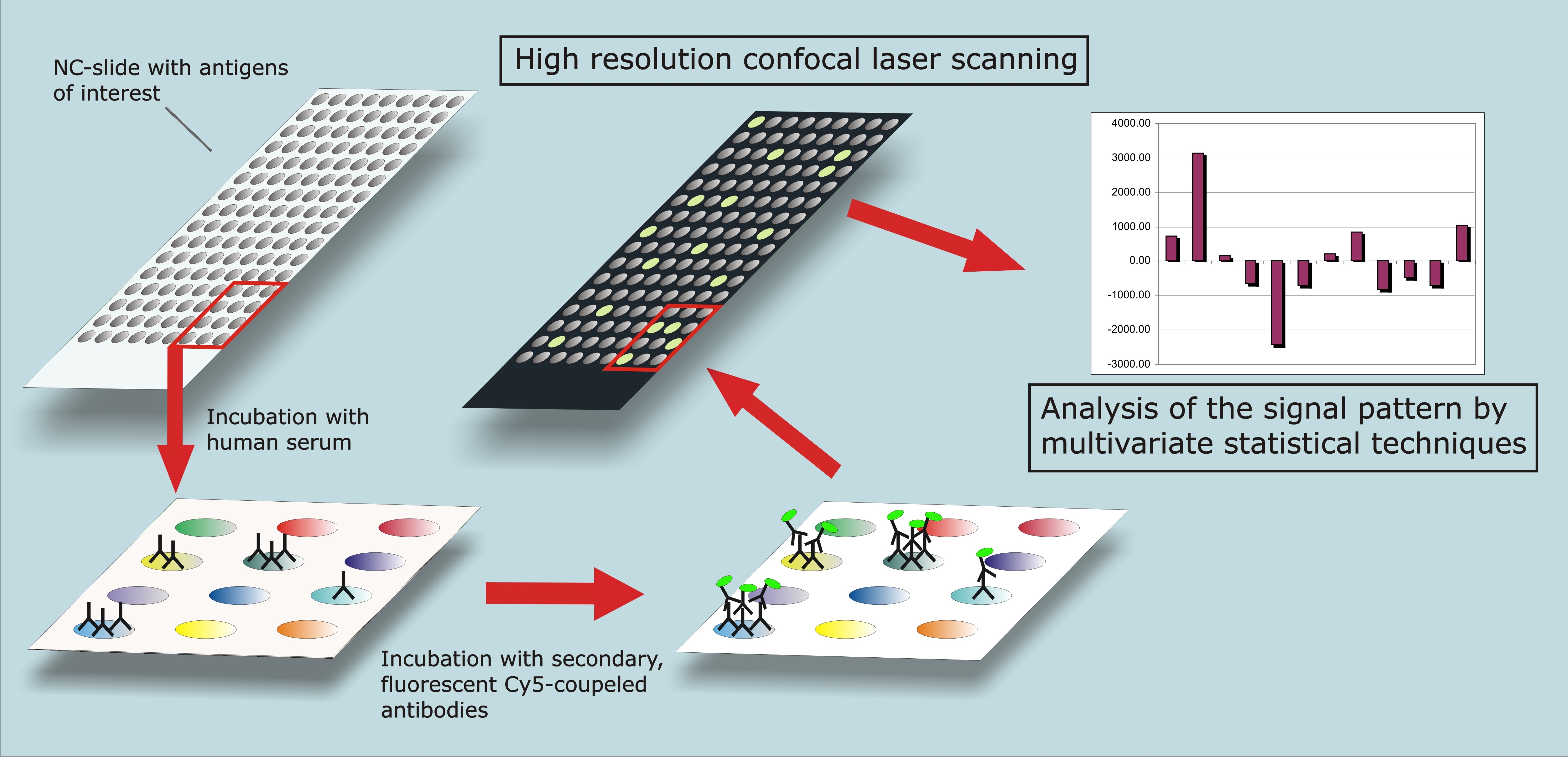
Fig. 1: Workflow and principle of protein microarray technology.
Protein microarrays
In the approach we use, patients’ body fluids, such as sera, tear fluid or aqueous humor are screened for subsets of antibodies against specific antigens of interest, in order to detect potential, disease related differences in antibody-antigen interactions (see fig.1). The protein array slides are prepared by spotting highly purified antigens onto special nitrocellulose-coated slides. Due to the high spotting density hundreds of different antigens can be tested on each customized protein microarray slide, allowing large scale screening studies. In order to capture antigen-specific antibodies, the arrays are incubated with the body fluids. Then the arrays are treated with a secondary fluorescence labeled antibody (Cy3, Cy5) for visualizing the antibody-antigen interactions. Subsequently, slides are scanned with a high-resolution-convocal-scanner whereby the fluorescence signals emitting from the secondary antibodies are digitized. In order to compare spot signals and to detect potential biomarkers we are using a combination of different algorithms for data normalization and a set of diverse statistical techniques, such as artificial neural networks, multivariate statistics and tree algorithms.
Antibody microarrays
In this approach antibodies instead of antigens are spotted onto nitrocellulose coated slides and the arrays are incubated with patients’ body fluids for capturing antigens of interest. For the purpose of using a high resolution convocal laser scanner, proteins are labeled with a fluorescence dye (Cy3, Cy5) prior to the incubation with the body fluid. The subsequent data analysis and the statistical evaluation are based on the same algorithms and techniques as described for protein microarrays.The advantage of this technique is a rapid confirmation of potential biomarkers obtained from other clinical studies in a high throughput compatible manner. Furthermore it is possible to flexibly screen for high quantities of putative biomarkers.