Cell Culture
Possibilities of cell culture and proteomics
Working with cell culture is not only an established model for many diseases but also offers a quick and efficient way of analyzing the reaction and response of various types of cells on different effects or conditions. In our case this includes not only looking at the viability of the cells but also much deeper at the proteomics of the cells. The advantage of using cell culture is the high throughput and reproducibility throughout the different studies given through immortalized cell lines. Therefore cell culture is widely used to understand diseases and drug effects on cells.
Retinal Ganglion cells:
Representing a group of ocular diseases glaucoma is one of the leading causes of blindness. It leads to a progressive loss of retinal ganglion cells and visual field. Although this disease is wide spread the cause is still to be found. Although retinal ganglion cells are one of the important targets in glaucoma little is known about the cells reactions to the disease. Using different retinal ganglion cell lines we are trying to understand the pathogenesis of the disease. Most types of glaucoma are associated with an elevation of the intraocular pressure. Several studies have been able to show apoptosis of retinal ganglion cells by putting them under an elevated hydrostatic pressure. Our aim is to take a deeper look into the procedures happening in the cells by looking at the proteomics of the cells after exposing them to different pressure conditions. In addition our group could show the presence of antibodies against ocular antigens in sera of patients suffering of glaucoma. Therefore, using two different immortalized retinal ganglion cell lines, we are also investigating the effects of sera on the viability as well as the proteomics of retinal ganglion cells.
The aim of these studies is to better understand the disease Glaucoma. By analyzing the proteomic reaction of the cells we furthermore are trying to find specific proteins that could serve as possible drug targets.
Literature:
Bell K, Rosignol I, Sierra-Filardi E, Rodriguez-Muela N, Schmelter C, Cecconi F, Grus F, Boya P (2020) Age related retinal Ganglion cell susceptibility in context of autophagy deficiency. Cell Death Discov 6:21.
Bell K, Wilding C, Funke S, Perumal N, Beck S, Wolters D, Holz-Muller J, Pfeiffer N, Grus FH (2016) Neuroprotective effects of antibodies on retinal ganglion cells in an adolescent retina organ culture. J Neurochem 139:256-269.
Wilding C, Bell K, Funke S, Beck S, Pfeiffer N, Grus FH (2015) GFAP antibodies show protective effect on oxidatively stressed neuroretinal cells via interaction with ERP57. J Pharmacol Sci 127:298-304.
Bell K, Wilding C, Funke S, Pfeiffer N, Grus FH (2015) Protective effect of 14-3-3 antibodies on stressed neuroretinal cells via the mitochondrial apoptosis pathway. BMC Ophthalmol 15:64.
Wilding C, Bell K, Beck S, Funke S, Pfeiffer N, Grus FH (2014) gamma-Synuclein antibodies have neuroprotective potential on neuroretinal cells via proteins of the mitochondrial apoptosis pathway. PLoS One 9:e90737.
Bell K, Funke S, Pfeiffer N, Grus FH (2012) Serum and antibodies of glaucoma patients lead to changes in the proteome, especially cell regulatory proteins, in retinal cells. PLoS One 7:e46910.
Results have been presented:
ARVO 2012/ 2013 -
Bell K, Wilding C, Funke S, Pfeiffer N, Grus F (2013) Changes in antibodies from glaucoma patients lead to changes in apoptosis pathways of neuroretinal cells. Invest Ophthalmol Vis Sci 54:1605-.
Wilding C, Bell K, Funke S, Beck S, Pfeiffer N, Grus F (2013) Protective effect of GFAP antibody on RGC5 cells via actin cytoskeleton pathways. Invest Ophthalmol Vis Sci 54:432-.
Bell K, Wilding C, Pfeiffer N, Grus FH (2012) Down Regulation Of 14-3-3 Ab In Glaucoma Patients Could Lead To Loss Of Protective Effects. Invest Ophthalmol Vis Sci 53:6592-.
Wilding C, Bell K, Grus F, Pfeiffer N (2012) Effect Of {gamma}- Synuclein Antibody On Rgc5 And Mitochondrial Apoptosis Pathways. Invest Ophthalmol Vis Sci 53:6591-.
Ophthalmopharmacology:
Keeping in mind that treatment of glaucoma still is not at its perfection we are not only interested in the reaction of the cells towards pressure but also towards different pharmaceutical drugs. Glaucoma, as a progressive disease leading to apoptosis of retinal ganglion cells, can be looked at as a neurodegenerative disease. Therefore our group is especially interested in finding neuroprotective agents that protect the cells against this neurodegeneration. After knowing the proteomic reaction of the retinal ganglion cell towards pressure one of our next aims is to investigate if there are substances to be found that can prevent this reaction of the cells and if these proteomic biomarkers can serve as innovative therapeutic treatment options. Previous studies have been able to show an involvement of COX-II in other neurodegenerative diseases and also were able to show a neuroprotective Effect of COX-II Inhibitors. As Glaucoma also is a neurodegenerative disease we the idea arouse to investigate these substances in combination with glaucoma. Therefore working together with the Institute of Pharmacy (PD Dr. H. Ulbrich) we have been analyzing the effects of COX-I and COX-II inhibitors on retinal ganglion cells in order to see if they also have a positive effect on retinal ganglion cells. In first studies we could show a neuroprotective effect of both COX-I and COX-II inhibitors. In addition we could find that COX-II inhibitors were the more potent neuroprotective agents. This was demonstrated by less loss of viability under an elevated pressure as well as less proteomic reaction of the cells towards an elevated pressure.
Literature:
Brust AK, Ulbrich HK, Seigel GM, Pfeiffer N, Grus FH (2008) Effects of Cyclooxygenase Inhibitors on Apoptotic Neuroretinal Cells. Biomark Insights 3:387-402.
Ocular surface diseases, contact lenses:
One of the most frequent ocular surface diseases is the dry eye syndrome. This disorder is also very common among contact lens wearers but can also be caused by various medications or occur in a very high percentage of patients following refractive surgery e.g. LASIK operation. Belonging to the ocular surface is the cornea and the conjunctiva. In our group we work with conjunctival and corneal cells. One hypothesis about the dry eye disease in contact lens wearers is that it is the long term reaction of the ocular surface towards the different contact lens solutions used to clean the contact lenses. In previous studies analyzing tears we were able to demonstrate changes in its protein profiles depending on the used contact lens solution. These results lead to the conclusion that the contact lens solutions not only affect the tears but also the cells they get in contact with. The direct effects of these solutions on the ocular surface cells are still unknown. Using corneal and conjunctival cells we are starting to investigate if and what effects can be seen.
Literature:
Bell K, Buksinska E, Pfeiffer N, Grus FH (2012) Comparison of the effects of different lens-cleaning solutions on the protein profiles of human conjunctival cells. Graefes Arch Clin Exp Ophthalmol 250:1627-1636.
Kramann C, Boehm N, Lorenz K, Wehrwein N, Stoffelns BM, Pfeiffer N, Grus FH (2011) Effect of contact lenses on the protein composition in tear film: a ProteinChip study. Graefes Arch Clin Exp Ophthalmol 249:233-243.
Proteomics:
In our laboratory we are not only interested in the obvious effects on the cells such as viability but want to take a much deeper look into the cells by analyzing the whole protein expression profiles. In our studies we analyze the proteins in order to find specific biomarkers. These then can be identified and possibly can be used as new drug target. Due to the fact that our laboratory is equipped to a very high standard we are able to work with basic proteomic methods such as gel electrophoresis and HPLC but also with methods such as Seldi- Tof- Ms as well as Maldi- Tof –Tof- MS.
Depending on the study the workflow after creating a cell-lysate sample can vary. Protein profiles can be directly measured by Seldi-Tof-MS and Maldi-Tof-Tof-MS.
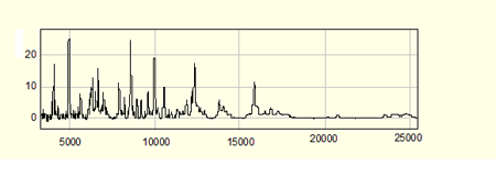
This is an example of a Seldi-Tof-MS measurement of a protein profile from a retinal ganglion cell. The X-Axis shows the molecular weight of the measured proteins, the Y- Axis shows the intensity of the protein in the sample.
The protein profiles are very complex. Therefore to take an even deeper look at the procedures in the cells the samples can be pre-fractioned by SDS-Page, HPLC or beads. The fractions then can be measured with mass spectrometry and also digested. The digested proteins then can be identified by Maldi-Tof-Tof-MS. All of our studies undergo complex statistic analysis which also -but not only- provides us with the most significant biomarkers. Our aim is to especially identify these proteins.
FACS (Fluorescence activated cell sorting):
To measure the survival respectively the apoptosis of the cells after the experiment we have different methods available. One very common test we also use is the colorimetric WST-1 Test. This is a formazan-based test which shows the surviving cells. Of course this test can only give an overview of the happenings in the sample.
Therefore much more accurate tests are needed to be able to analyze apoptosis of the cells. In our laboratory we have a FACS available. With this method we are able to analyze the exact apoptosis and necrosis of the cells during the experiment. The cells are incubated with different fluorescent markers such as Annexin-V and Propidiumiodid that then can be detected. The machine can count the marked cells and therefore analyze both apoptosis and necrosis. The advantage of this method in comparison to other methods is that it shows not only the cells that are in apoptosis in the very moment of the measurement but can also show those cells that have already gone through apoptosis.
The FACS Machine can not only be used to analyze apoptosis. It is a very wide spread method to analyze the expression of single proteins or receptors in or on the cells which can provide us with further valuable information.