Immunoproteomics profiling
The up- and down regulation of proteins can provide important informations about disease processes and can lead to a better understanding of the pathogenesis. This process might identify important biomarkers of the disease and thus will lead to predictive medicine.
Beside conventional two-dimensional electrophoresis, our group is using the Seldi-TOF (surface enhanced laser desorption and ionization in time-of-flight mass spectrometry) ProteinChip. Furthermore, we have state-of-the technologies in our proteomics unit in our lab. This includes a Maldi-TOFTOF (Bruker Ultraflex), different robots for sample preparation, HPLC and nano-HPLC systems coupled to automated Maldi-Target spotting, and Protein Microarray.
Recently, we could use this technology to find biomarkers for dry-eye disease (s. News section).
Bead Approach
Previous studies on antibodies in glaucoma showed that there exist complex IgG antibody repertoires and significant different patterns of antibodies against ocular antigens between glaucoma patients and normal subjects, using Western blotting.
Immunodetection after Western Blotting can effectively detect specific antibody reactivity in a given sample. However, this technique is time consuming, it is hard to eliminate blot-to-blot variation, and it has a limitation in sensitivity when used for mass screening of many samples simultaneously. In comparison, a bead-based assay could capture immunoglobulins for downstream immunoprecipitation of a wide range of proteins (from a large amount of samples) or other antigens, such as retina components, in a short period of time. This method has less variation, and is more sensitive, since proteins are detected by mass spectrometry (MS). Therefore, we are also working with a bead-based antibody assay (figure 1).
Figure 1: Protein G bead experiment: magnetic Protein G beads were incubated with serum samples in order to bind serum antibodies to the bead. These complexes were incubated with the retinal antigens. The bound antigens were eluted through pH shift and the elutions were measured by mass spectrometry.
Protein Microarrays
Western Blotting
Aim of one of our current studies is to analyze the complex IgG autoantibody patterns against human retinal and optic nerve antigens in patients with glaucoma. Since previous studies used mainly bovine antigens, we are trying to show that the different antibody profiles between glaucoma patients and healthy subjects are truly autoantibodies and the complex profiles are not only based on cross-reactivities with non-human antigens.
Serum samples from several groups will be analyzed in this project:
• Non-glaucomatous healthy control subjects (CO)
• Primary open-angle glaucoma subjects (POAG)
• Subjects with ocular hypertension (OHT)
• Normal tension glaucoma patients (NTG)
Figure 2. Examples of Western blots made visible by 4-chloro-1-naphtol staining. There are complex IgG autoantibody patterns in all four groups: healthy subjects (CO), patients with primary open-angle glaucoma (POAG), ocular hypertension (OHT), and normal tension glaucoma (NTG). Each lane was incubated with patient serum (diluted 1:40) as first antibody and goat anti-human IgG (diluted 1:500) as secondary antibody.
Figure 3 shows the synopsis of complex antibody profiles for all three groups. The mean antibody reactivity is plotted against the corresponding molecular weight (in kDa). A significant difference can be seen between autoantibody profiles of the POAG and the CO group (P=0.0040, Mahalanobis distance=1.03), between the NTG group and the control group (P=0.000035, Mahalanobis distance=1.63), and also between the POAG and NTG group (P=0.021).
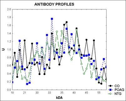
Figure 3: Synopsis of antibody profiles against human optic nerve antigens. The mean antigen-antibody-reactivity of patients with primary open-angle glaucoma (POAG), normal tension glaucoma (NTG), and control subjects (CO) is plotted against the corresponding molecular weight of the optic nerve antigens. The x-axis shows the molecular weight of the antigen-antibody-reactivity in Kilodalton (kDa).
We could detect complex IgG autoantibody repertoires against human optic nerve antigens (see figure 2) and significant differences (P<0.05) could be shown among the profiles of all groups (POAG, NTG, and CO). In accordance with studies using bovine optic nerve antigens we did not only find up-regulated antibody reactivities in the glaucoma groups of this project, but also down-regulated areas.
At around 16 kDa a significant difference between the antibody reactivity of the three groups could be seen (P=0.029). The POAG and the NTG group showed an up-regulation in this area in comparison to the control group. The corresponding antigen was identified as myelin basic protein (MBP). MBP is a trans-membrane protein which plays an important role in the myelination process in the central and peripheral nervous system.
Recent publications
-
Korb CA, Gerstenberger E, Lorenz K, Bell K, Beck A, Scheller Y, Beutgen VM, Wolters D, Grus FH (2024) Correlation of Functional and Structural Outcomes with Serum Antibody Profiles in Patients with Neovascular Age-Related Macular Degeneration Treated with Ranibizumab and Healthy Subjects: A Prospective, Controlled Monocenter Trial. Journal of clinical medicine 13.
-
Korb CA, Beck S, Wolters D, Lorenz K, Pfeiffer N, Grus FH (2023a) Serum Autoantibodies in Patients with Dry and Wet Age-Related Macular Degeneration. Journal of clinical medicine 12.
-
Korb CA, Lackner KJ, Wolters D, Schuster AK, Nickels S, Beutgen VM, Munzel T, Wild PS, Beutel ME, Schmidtmann I, Pfeiffer N, Grus FH (2023b) Association of autoantibody levels with different stages of age-related macular degeneration (AMD): Results from the population-based Gutenberg Health Study (GHS). Graefe's archive for clinical and experimental ophthalmology = Albrecht von Graefes Archiv fur klinische und experimentelle Ophthalmologie.
- Beutgen VM, Pfeiffer N, Grus FH (2021) Serological Levels of Anti-clathrin Antibodies Are Decreased in Patients With Pseudoexfoliation Glaucoma. Frontiers in immunology 12:616421.
- Beutgen VM, Schmelter C, Pfeiffer N, Grus FH (2020) Autoantigens in the trabecular meshwork and glaucoma-specific alterations in the natural autoantibody repertoire. Clin Transl Immunology 9:e01101.
- Liu H, Anders F, Funke S, Mercieca K, Grus F, Prokosch V (2020) Proteome alterations in aqueous humour of primary open angle glaucoma patients. International journal of ophthalmology 13:176-179.
- Schmelter C, Fomo KN, Perumal N, Manicam C, Bell K, Pfeiffer N, Grus FH (2019) Synthetic Polyclonal-Derived CDR Peptides as an Innovative Strategy in Glaucoma Therapy. J Clin Med 8.
- Beutgen VM, Perumal N, Pfeiffer N, Grus FH (2019) Autoantibody Biomarker Discovery in Primary Open Angle Glaucoma Using Serological Proteome Analysis (SERPA). Front Immunol 10:381.
- Bell K, Funke S, Grus FH (2019) [Autoimmunity and glaucoma]. Ophthalmologe 116:18-27.
- Schmelter C, Perumal N, Funke S, Bell K, Pfeiffer N, Grus FH (2017) Peptides of the variable IgG domain as potential biomarker candidates in primary open-angle glaucoma (POAG). Hum Mol Genet 26:4451-4464.